If linker peptide is inhibited, the monomers are not linked and the cell wall is not synthesized.

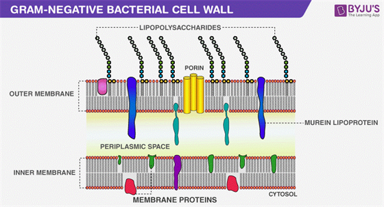
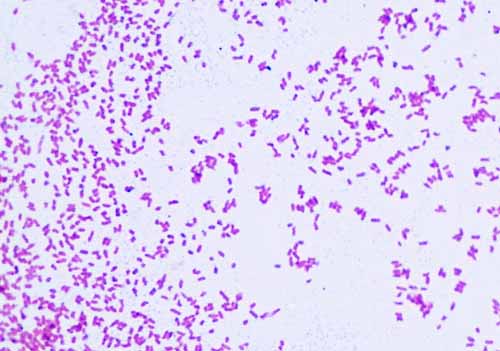
Gram-positive bacteria have a high content of peptidoglycan that is present in their cell wall. They have a thin layer of peptidoglycan that is present in the cell wall. Gram-negative bacteria have a low content of peptidoglycan. These polymers combine to give the structure of the bacterial cell wall. These amino acids are homopeptide and are similar to each other. Three to five peptide chains linked the sugar and protein together. The carbohydrates that are N-acetylglucosamine and N- acetylmuramic acid are linked with the amino acids. This peptidoglycan is the polymers of sugar and amino acids. They are the complex polysaccharides that synthesize the cell wall of bacteria. Peptidoglycan is the monomeric form of carbohydrate that is only found in bacteria only. There are three components of the gram-negative bacterial cell wall. It is a two-layered cell wall that has 20-30% lipid content and is 10-20% murein content. They have a single layer of peptidoglycan. Gram negative bacterial cell wall has some differences than the gram-positive cell wall. Because of the change in cell wall stricture, the bacteria are classified as gram positive bacteria and gram negative bacteria. This assay can be integrated into a simple diagnostic platform for rapid screening tests and the stratification of Gram-negative bacterial infections in the clinic.Gram-negative bacteria have different cell wall than Gram positive bacteria. Further, we determined that our method can be used for complex samples, such as combinations of multiple bacterial types bacteria in the presence of mammalian cells and bacteria with serum components.


By using the COL-FL assay, we achieved the differential labeling of various Gram-negative pathogens related to hospital-acquired infections, which could be subsequently detected via spectrofluorometry and microscopy. Herein, we introduce a method utilizing a fluorescent derivative of colistin (COL-FL), that can directly label the Gram-negative cell wall of live bacteria and universally detect the targets within 10 min. A rapid and simple diagnostic method would enable immediate and targeted treatment, while dramatically reducing antibiotic overuse. Currently, a Gram-negative infection is diagnosed by symptom evaluation and is treated with empiric antibiotics which target both Gram-negative and Gram-positive bacteria. Gram-negative bacteria are a significant cause of infections acquired in both hospital and community settings, resulting in a high mortality rate worldwide.


 0 kommentar(er)
